WunderkanoneExploring the Microscopic World
Nuclearia
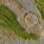
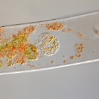
Spirogyra cell overtaken by Nuclearia sp..
Spirogyra cell overtaken by Nuclearia sp..
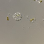
Resting cell during phagocytosis. The hair-like corona are bacteria that feed on the muccous layer surrounding the Nuclearia.
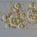
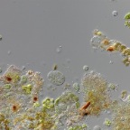
Different types of forms from left to right: resting cell during phagocytosis, cyst, resting cell during phagocytosis, hungry cell looking for a Spirogyra to invade. The cyst is surrounded by a muccous layer as well. The brown particals in this layer are rests of phagocytosis.
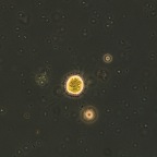
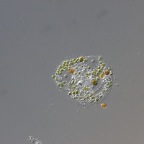
Cyst
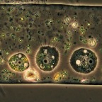
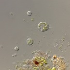
Can you count the number of Nuclearia sp. in this image? In the upper right corner there is a Pseudospora parasitica zoocyst visible.
Nuclearia sp.
Nuclearia sp.
Nuclearia sp.
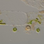
Nuclearia sp. from another sample with Spirogyra algae. This could be N. delicatula.